|
Betula kenaica |
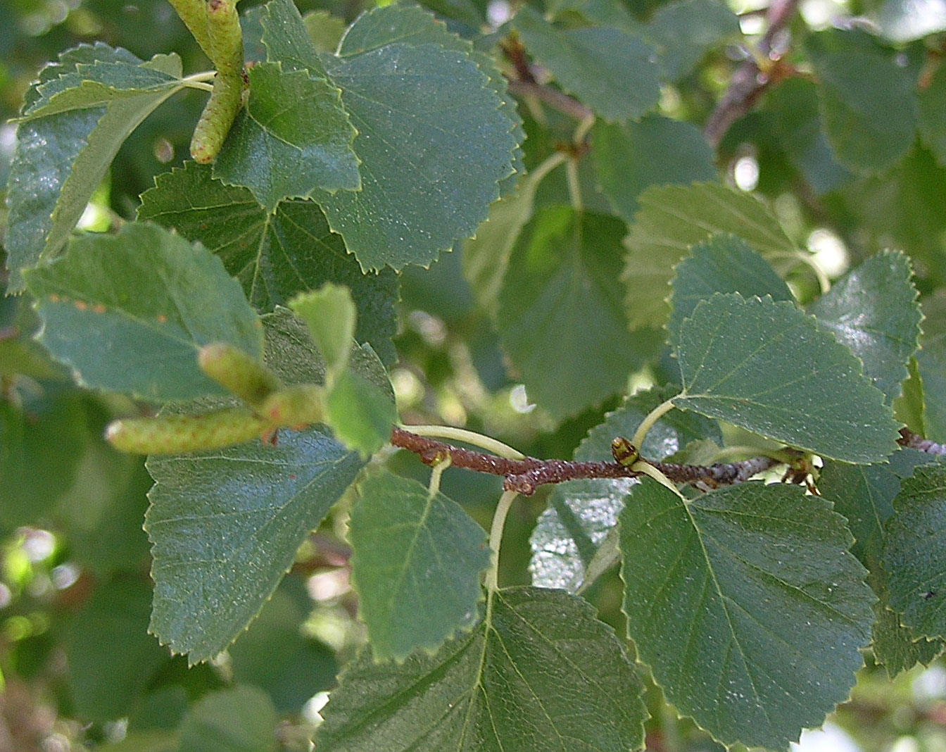
Betula kenaica
|
|
Betula kenaica
|
Betula nana
|
|
Betula occidentalis |
|
| Buruk K., A. Sokmen, F. Aydin and M. Erturk. 2006. Antimicrobial activity of some endemic plants growing in the Eastern Black Sea Region, Turkey. Fitoterapia 77(5):388–391. “The Eastern Black Sea Region has an extensive flora because of ample rainfall lasting all year. In this study, antimicrobial effects of 74 crude extracts of 22 endemic plants were investigated. Among the 30 active crude extracts, water-insoluble crude extracts from Betula medwediewii, Heracleum platytaenium, Primula longipes, Anthemis cretica ssp. argaea and Centaurea helenioides were the prominent ones with their MIC values.” Gong Y., R. M. Raj, C. A. Luscombe, I. Gadawski, T. Tam, J. Chu, D. Gibson, R. Carlson and S. L. Sacks. 2004. The synergistic effects of betulin with acyclovir against herpes simplex viruses. Antiviral Res. 64(2):127–130. “Betulin, a pentacyclic triterpenoid, was isolated from the bark of Betula papyrifera. The antiviral efficacies of betulin on herpes simplex virus type 1 (HSV-1) and type 2 (HSV-2) were evaluated using viral plaque reduction assays on Vero cells. The results indicate that betulin is active against both HSV-1 and HSV-2 infections with the 50% effective concentrations (EC(50)) of 0.40 and 4.15 microg/ml, respectively. The cytotoxicity of betulin was examined on Vero cells using a neutral red uptake assay. The 50% cytotoxic concentration (CC(50)) of betulin was 73.1 microg/ml. A synergistic antiviral effect between betulin and acyclovir (ACV) was determined by drug combination studies. Strong and moderate synergistic antiviral effects were observed for betulin and ACV against HSV-1 when the concentrations of ACV and betulin were higher than 0.068 and 0.4 microg/ml, respectively. At the concentrations lower than these, additive effect was found. Synergistic antiviral effects were also found against HSV-2 at higher concentrations than for HSV-1, i.e. 0.45 microg/ml of ACV combined with 8.4 microg/ml of betulin.” Huyke C, M. Laszczyk, A. Scheffler, R. Ernst and C. M. Schempp. 2006. Treatment of actinic keratoses with birch bark extract: a pilot study. J. Dtsch. Dermatol. Ges. 4(2):132–136. In German. “BACKGROUND: Birch bark contains a variety of apoptosis-inducing and anti-inflammatory substances such as betulinic acid, betulin, oleanolic acid and lupeol. Therefore, birch bark extract may be effective in the treatment of actinic keratoses. To address this issue, a pilot study using a standardized birch bark ointment was performed. METHODS: Twenty-eight patients with actinic keratoses were enrolled in this prospective, non-randomized pilot study. Fourteen patients were treated with birch bark ointment only; fourteen patients received a combination therapy with cryotherapy and birch bark ointment. Treatment response was assessed clinically after two months. RESULTS: Clearing of more than 75 % of the lesions was seen in 79 % of the patients treated with birch bark ointment monotherapy. The response rate of the combined treatment modality was 93 %. Therapy with birch bark ointment was well tolerated. CONCLUSION: In this pilot study, a standardized birch bark extract was effective in the treatment of actinic keratoses. This therapy is easy to perform and it has no side effects. Birch bark ointment may be a new therapeutic option for actinic keratoses.” Ju E. M., S. E. Lee, H. J. Hwang and J. H. Kim. 2004. Antioxidant and anticancer activity of extract from Betula platyphylla var. japonica. Life Sci. 74(8): 1013–1026. “The antioxidant and anticancer properties of a medicinal plant, Betula platyphylla var. japonica were investigated. The total methanol extract of B. platyphylla var. japonica had protective effects against hydrogen peroxide (H2O2) in the Chinese hamster lung fibroblast (V79-4) cell line and induced apoptotic cell death in human promyelocytic leukemia (HL-60) cells, a cancer cell line. B. platyphylla var. japonica extract significantly increased cell viability against H2O2. The extract also showed high 1,1-diphenyl-2-picrylhydrazyl (DPPH) radical scavenging activity (IC50 2.4 microg/ml) and lipid peroxidation inhibitory activity (IC50 below 4.0 microg/ml). Furthermore, B. platyphylla var. japonica extract reduced the number of V79-4 cells arrested in G2/M in response to H2O2 treatment and increased the activities of several cellular antioxidant enzymes, including superoxide dismutase, catalase and glutathione peroxidase. Treatment with B. platyphylla var. japonica extract induced cytotoxicity and apoptosis in HL-60 cells, as shown by nucleosomal DNA fragmentation, increases in the subdiploid cell population, and fluorescence microscopy. B. platyphylla var. japonica extract gradually increased the expression of pro-apoptotic Bax and led to the activation of caspase-3 and cleavage of PARP. These findings suggest that B. platyphylla var. japonica exhibits potential antioxidant and anticancer properties.” Kashiwada Y., M. Sekiya, K. Yamazaki, Y. Ikeshiro, T. Fujioka, T. Yamagish, S. Kitagawa and Y. Takaishi. 2007. Triterpenoids from the floral spikes of Betula platyphylla var. japonica and their reversing activity against multidrug-resistant cancer cells. J. Nat. Prod. 70(4): 623–627. “Four new triterpenes, together with 16 known triterpenes, were isolated from the floral spikes of Betula platyphylla var. japonica in a search for compounds capable of reversing multidrug resistance in cancer cells. The structures of the new triterpenes were elucidated as 3,4-seco-olean-4(23),13(18)-dien-3-oic acid (1), 3,4-seco-urs-4(23),20(30)-dien-3-oic acid (2), 3-O-methylmalonylepiocotillol II (6), and 3-O-methylmalonylcabraleahydroxylactone (16) by spectroscopic examination. The cytotoxicity of the isolated triterpenes against human cancer cell lines as well as multidrug-resistant cancer cell lines was evaluated. Most of the isolated triterpenes showed very weak cellular toxicities. Although no discernible differences were found in the cytotoxicities for the tested compounds against sensitive and resistant cell lines, the cytotoxicities for several triterpenes against multidrug-resistant cancer cell lines (KB-C2 or K562/Adr) were enhanced in the presence of nontoxic concentrations of colchicine or doxorubicin. Compound 10 reversed the cytotoxicity of colchicine against KB-C2 cells at 8.1 microM and showed comparable potency to 5 microM verapamil.” Laitinen J., R. Julkunen-Tiitto, M. Rousi, J. Heinonen and J. Tahvanainen. 2005. Ontogeny and environment as determinants of the secondary chemistry of three species of white birch. J. Chem. Ecol. 31(10): 2243–2262. “This study investigates variation in the secondary chemistry of the bark of three closely related, winter-dormant species of white birch (Betula resinifera, B. pendula, and B. platyphylla) at different ontogenetic stages by using different plant parts (top and base). The experimental birches were grown for 4 years in two growing conditions (pot and field) at different nutrient levels. There was considerable species-specific quantitative and qualitative variation in the secondary chemicals in bark, but this was also affected by fertilization and the age of the plant. In general, there was greater chemical diversity in saplings than in seedlings. The study revealed three new components, secoisolariciresinol 9-O-beta-glucopyranoside and two of its derivatives, that have not been reported previously for the bark of white birches. Principal component analysis showed that the species studied had a similar chemical composition at the juvenile stage, but as the plants grew, they became more clearly differentiated, which indicates that the species of older plants can be identified by chemotaxonomy. Evidently, the secondary chemistry of birches is under genetic control, but it is affected by properties of growing conditions and ontogeny.” Laszczyk M., S. Jager, B. Simon-Haarhaus, A. Scheffler and C. M. Schempp. 2006. Physical, Chemical and Pharmacological Characterization of a New Oleogel-Forming Triterpene Extract from the Outer Bark of Birch (Betulae Cortex). Planta Med.: “Triterpenes are biologically active secondary plant substances that display antimicrobial, hepatoprotective and anti-inflammatory effects. However, the poor solubility of triterpenes in both polar and non-polar solvents as well as expensive purification procedures have prevented the large-scale isolation of these compounds for medicinal purposes. Here, we describe a novel quantitative extraction method of triterpenes from the outer bark of birch ( BETULA species) in which betulin, a lupan triterpene, predominates. The resulting highly purified triterpene extract (TE) in the form of a dry powder contains betulin as the major compound, but also betulinic acid, lupeol, erythrodiol and oleanolic acid. We have found that this TE is able to form an oleogel, thus providing an opportunity for the topical application of pharmacologically relevant amounts of triterpenes. Furthermore, we have investigated the TE in comparison to its major isolated compounds in cell culture experiments with human immortalized keratinocytes and skin cancer cells. We could demonstrate dose-dependent cytotoxic and apoptosis-inducing effects of TE and betulin. These experimental data support the notion from a previous clinical study that TE from the outer bark of birch might represent a new tool for the topical treatment of skin cancer and skin cancer precursors like actinic keratoses.” Mshvildadze V., J. Legault, S. Lavoie, C. Gauthier and A. Pichette. 2007. Anticancer diarylheptanoid glycosides from the inner bark of Betula papyrifera. Phytochemistry 68(20): 2531–2536. “Phytochemical investigations of the MeOH extract of Betula papyrifera inner bark led to the isolation of ten phenolic compounds of the following types: diarylheptanoid glycosides (1-4), a diarylheptanoid (5), a lignan (6), flavonoids (7-8) and chavicol glycosides (9-10). Among them, the diarylheptanoid glycoside, (S)-1,7-bis-(4-hydroxyphenyl)-heptan-3-one-5-O-alpha-l-arabinofuranosyl-(1-->6)-beta-d-glucopyranoside, papyriferoside A (1), was isolated and its structure was determined on the basis of 1D and 2D NMR, HPLC-MS, as well as high resolution mass spectroscopic data. Platyphylloside (4) exerted the strongest cytotoxic activity of all isolated compounds with IC(50) values ranging from 10.3 to 13.8muM.” Sekiya M., Y. Kashiwada, T. Nabekura, S. Kitagawa, T. Yamagishi, T. Yokozawa, T. Ichiyanagi, Y. Ikeshiro and Y. Takaishi. 2007. Effect of Triterpenoids Isolated from the Floral Spikes of Betula platyphylla var. japonica on P-Glycoprotein Function. Planta Med. Dec. “One of the major causes of multidrug resistance (MDR) in cancer cells is over-expression of P-glycoprotein (P-gp). We studied the effects of 20 triterpenes isolated from the floral spikes of BETULA PLATYPHYLLA var. JAPONICA ( B. PLATYPHYLLA) on P-gp function based on our previous finding that some of them showed MDR reversing effects. We evaluated accumulations and effluxes of rhodamine 123 as a P-gp substrate with P-gp over-expressing KB-C2 cells. Among the 20 triterpenes, compounds 3, 4, 8, 9, 13, 15, and 20 increased rhodamine 123 accumulations in KB-C2 cells, and three ( 8, 13, and 20) of them also inhibited efflux of rhodamine 123 out of cells. In addition, compounds 13 and 20 showed a weak inhibitory activity of P-gp ATPase. These results suggested that MDR reversing effects of compounds 13 and 20 are partly involved in inhibition of P-gp ATPase. B. platyphylla: BETULA PLATYPHYLLA var. JAPONICADMEM:Dulbecco's modified Eagle's medium EDTA:ethylendiamine- N, N, N', N'-tetraacetic acid EGTA: O, O'-bis(2-aminoethyl)ethyleneglycol- N, N, N', N'-tetraacetic acid FBS:fetal bovine serum HEPES:2-[4-(2-hydroxyethyl)-1-piperazinyl]ethane-sulfonic acid MDR:multidrug resistance PBS:phosphate-buffered saline P-gp:P-glycoprotein Tris:2-amino-2-hydroxymethyl-1,3-propanediol.” Thibeault D., C. Gauthier, J. Legault, J. Bouchard, P. Dufour and A. Pichette. 2007. Synthesis and structure-activity relationship study of cytotoxic germanicane- and lupane-type 3beta-O-monodesmosidic saponins starting from betulin. Bioorg. Med. Chem. 15(18): 6144-6157. “Germanicane-type triterpenes allobetulin (3) and 28-oxoallobetulin (4) can be obtained by the Wagner-Meerwein rearrangement of the more available lupane-type triterpenes betulin (1) and betulinic acid (2), respectively. The medical uses of betulinic acid (2) and its derivatives are limited because of their poor hydrosolubility and pharmacokinetics properties. In order to overcome this major problem, we synthesized and studied the in vitro anticancer activity of a series of 3beta-O-monodesmosidic saponins derived from betulin (14-16), betulinic acid (20-22), allobetulin (23-28) and 28-oxoallobetulin (29-34) based on six different natural sugar residues (d-glucose, l-rhamnose, d-arabinose, d-galactose, d-mannose and d-xylose). This structure-activity relationship study confirmed that betulinic acid saponins are generally better in vitro anticancer agents than those derived from betulin with the exception of betulin 3beta-O-alpha-d-mannopyranoside (15) which exerted a potent cytotoxic activity against lung carcinoma (A-549) and colorectal adenocarcinoma (DLD-1) human cell lines with IC(50) ranging from 7.3 to 10.1mumol/L. Furthermore, although the synthesis of novel germanicane-type saponins was carried out with success, the bioactivity measured for these glycosides was not as high as we anticipated since only the 3beta-O-beta-d-glucopyranoside and 3beta-O-beta-d-galactopyranoside of allobetulin (23,24) showed moderate anticancer activity (IC(50) 30-40 micromol/L).” |
|