|
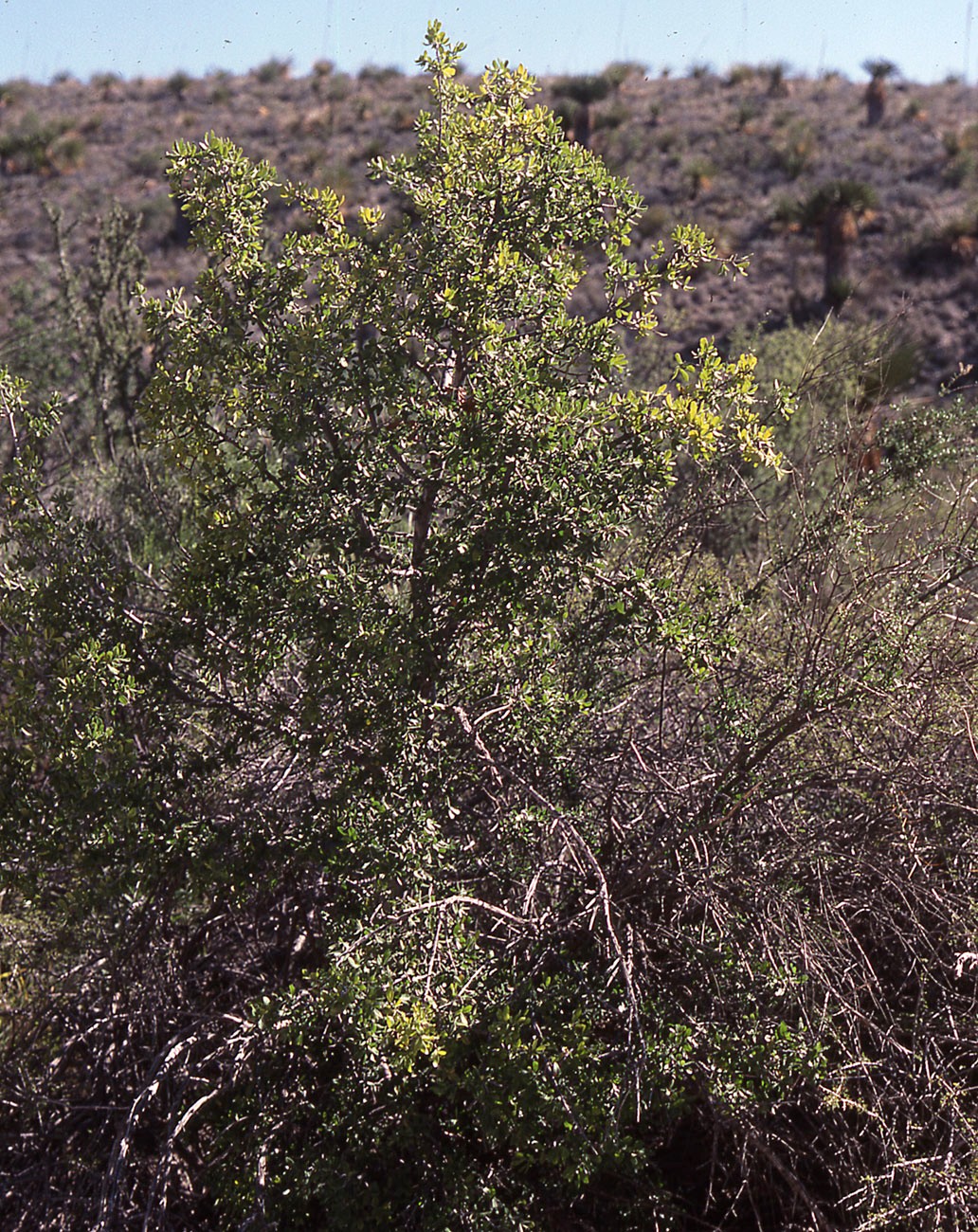
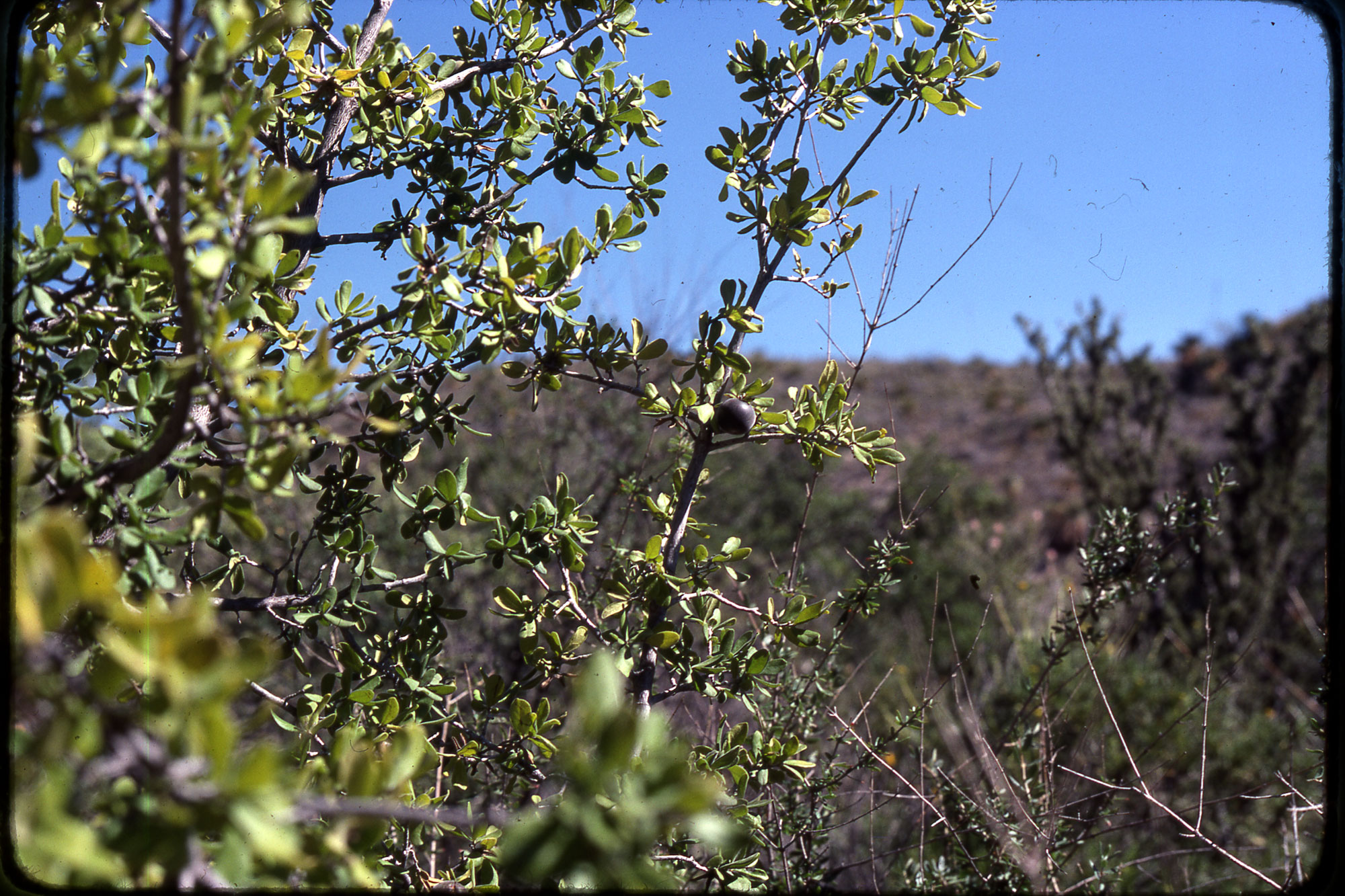
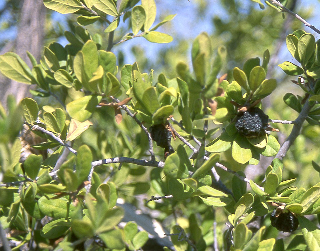
Diospyros texana |
Diospyros texana |
|
Achiwa Y., H. Hibasami, H. Katsuzaki, K. Imai and T. Komiya. 1997. Inhibitory effects of persimmon (Diospyros kaki) extract and related polyphenol compounds on growth of human lymphoid leukemia cells. Biosci. Biotechnol. Biochem. 61(7):1099–1101. “We have investigated the effects of persimmon (Diospyros kaki) extract (PS) and related polyphenol compounds such as catechin (C), epicatechin (EC), epicatechingallate (ECG), epigallocatechin (EGC), and epigallocatechingallate (EGCG) on the growth of human lymphoid leukemia Molt 4B cells. We found that PS, ECG, EGC, and EGCG strongly inhibited the growth of the cells in a dose-dependent manner, while C and EC inhibited the growth of the cells only moderately. Ornithine decarboxylase (ODC), a rate-limiting enzyme of polyamine biosynthesis, was inhibited by 10-20% by these polyphenol compounds. The morphology of the Molt 4B cells indicated severe damage 3 days after treatment with PS, ECG, EGC, and EGCG. Irregular shape of the cells and DNA fragmentation were observed in PS, ECG, EGC, or EGCG-treated cells. These results suggest that PS, ECG, EGC, and EGCG induce apoptosis (programmed cell death) of Molt 4B cells.” Adeniyi BA, Fong HH, Pezzuto JM, Luyengi L, Odelola HA. 2000. Antibacterial activity of diospyrin, isodiospyrin and bisisodiospyrin from the root of Diospyros piscatoria (Gurke) (Ebenaceae). Phytother. Res. 14(2): 112–117. “Two dimeric naphthoquinones, diospyrin and isodiospyrin, isolated from the root of Diospyros piscatoria (Gurke), a common ingredient in several folk medicines, have been shown to have a broad spectrum of antibacterial activity. The minimum inhibitory concentrations (MICs) of diospyrin against Streptococcus pyogenes ATCC 12344 and Streptococcus pneumoniae ATCC 33400 ranged from 1.56 to 50 microg/mL. While those against Salmonella choleraesuis serotype typhi (S. typhi), ATCC 6539 and Mycobacterium chelonae ATCC 19977 were between 25 and 100 microg/mL. Isodiospyrin was more active than its racemic isomer diospyrin. The MICs against Gram-positive bacteria ranged from 0.78 to 50 microg/mL. While those against Pseudomonas aeruginosa ATCC 15443 and S. typhi ranged from 50 to 100 microg/mL. The MIC for M. chelonae was between 6.25 and 25 microg/mL. MICs were found to increase with the concentration of cells used for the inoculum. The MICs for Bacillus subtilis ATCC 6633 increased up to the highest concentration of cells tested. The same phenomenon was observed on M. chelonae, but with better effect in the latter. The kinetics of bacteria studies against both B. subtilis and M. chelonae increases with increasing concentration of isodiospyrin tested. Two tetrameric forms of plumbagin were isolated. The naphthoquinone bisisodiospyrin, gave MIC values between 300 and 400 micro g/mL. The second, as yet unidentified tetramer, was not active at 500 micro g/mL.” Adzu B., S. Amos, I. Muazzam, U. S. Inyang and K. S. Gamaniel. 2002. Neuropharmacological screening of Diospyros mespiliformis in mice. J. Ethnopharmacol. 83(1-2):139–143. “The neuropharmacological activities of the aqueous extract of Diospyros mespiliformis stem bark were screened in mice. The extracts effect on pentobarbital-induced sleeping time, pentylenetetrazole induced seizure, spontaneous motor activity (SMA), exploratory behaviour, and rota-rod performance (motor coordination) were evaluated. The extract (100 and 200 mg/kg p.o.) produced a significant (P<0.05) prolongation of pentobarbital-induced sleeping time, and reduced the SMA and exploratory behaviour. The extract prolonged onset of the phases of seizure activity but did not protect mice against lethality induced by pentylenetetrazole. It also failed to affect the motor coordination test. These results suggest that the extract contained an agent with neuropharmacological activity that may be sedative in nature.” Borges-Argáez R., C. I. Canche-ChayI,L. M. Peña-Rodríguez, S. Said-Fernández and G. M. Molina-Salinas GM. 2007. Antimicrobial activity of Diospyros anisandra. Fitoterapia 78(5): 370–372. “Leaves, root and stem bark of Diospyros anisandra were screened against two strains of Mycobacterium tuberculosis, one resistant and one susceptible to antibiotics, using the microplate Alamar blue assay test. The lypophylic fractions of the root and bark showed significant inhibitory activity against both strains, with the hexane fraction of the bark showing the strongest activity (MIC 6.25 microg/ml) against the resistant strain and a significant antimicrobial activity against Staphylococcus aureus, Bacillus subtilis, Candida albicans, Aspergillus niger, and Colletotrichum gloeosporioides. The bioassay-guided purification of the bioactive hexane fraction resulted in the isolation and identification of the naphthoquinone plumbagin as one of the metabolites responsible for the biological activity.” Cai L, Wei GX, van der Bijl P, Wu CD. 2000. Namibian chewing stick, Diospyros lycioides, contains antibacterial compounds against oral pathogens. J. Agric. Food Chem. 48(3): 909–914. “The twigs of Diospyros lycioides, a plant commonly known as "muthala", are frequently used as chewing sticks for the cleaning of teeth by rural and urban people in Namibia. Preliminary studies showed that a methanol extract of D. lycioides inhibited growth of selected oral pathogens. Subsequent bioassay-guided fractionation led to the isolation of four novel bioactive naphthalene glycosides, diospyrosides A, B, C, and D (1-4), and two known bioactive naphthoquinones, juglone (5) and 7-methyljuglone (6). The structures of the new compounds were elucidated using spectroscopic techniques including 1D and 2D NMR. These compounds inhibited the growth of oral cariogenic bacteria (Streptococcus mutans and Streptococcus sanguis) and periodontal pathogens (Porphyromonas gingivalis and Prevotella intermedia) at minimum inhibitory concentrations ranging from 0.019 to 1.25 mg/mL. Juglone exhibited the strongest inhibitory activity among these compounds.” Cantrell C. L., M. A. Berhow, B. S. Phillips, S. M. Duval, D. Weisleder and S. F. Vaughn. 2003. Bioactive crude plant seed extracts from the NCAUR oilseed repository. Phytomedicine 10(4): 325–333. “Over four-hundred crude extracts from 202 plant species distributed among 131 plant families were evaluated for their bioactivity against brine shrimp (Artemia salina). Activity was determined for both the organic (CH2Cl2:MeOH) and aqueous extracts against A. salina in a 96 well-plate assay. Of the greater than four-hundred extracts tested, 21 organic and 6 aqueous extracts demonstrated potent cytotoxic activity (LC50 = < 100 microg/ml). Three of these organic extracts (Crateva religiosa, Diospyros dichrophylla, and Olax subscorpioidea) were chosen for chemical investigations due to their high activity and a lack of prior investigations. Chemical analysis of these extracts resulted in the isolation of oleanolic acid (1) and 4-epi-hederagenin (2) from C. religiosa, isodiospyrin (3) from D. dichrophylla, and santalbic acid (4) from O. subscorpioidea.” Chakrabarty S., M. Roy, B. Hazra and R. K.Bhattacharya. 2002. Induction of apoptosis in human cancer cell lines by diospyrin, a plant-derived bisnaphthoquinonoid, and its synthetic derivatives. Cancer Lett. 188 (1-2): 85-93. “Diospyrin, a bisnaphthoquinonoid natural product, and three synthetic derivatives have been tested for their action in four human cancer cell lines: acute myeloblastic leukemia (HL-60), chronic myelogenic leukemia (K-562), breast adenocarcinoma (MCF-7) and cervical epithelial carcinoma (HeLa). In cells grown in appropriate media several derivatives elicited cytotoxicity as assessed by Trypan Blue dye exclusion, 3-(4,5-dimethylthiazol-2-yl)-2,5-diphenyltetrazoliumbromide reduction and DNA synthesis. Diethyl ether derivative (D7) was most effective in this regard while the parent compound diospyrin (D1) was least active (D7>D3>D2>D1). D7 was not cytotoxic toward normal human lymphocytes, suggesting its action is specific for tumor cells. On microscopic examination D7-treated cells exhibited characteristic morphological features of apoptosis, such as cell shrinkage and formation of apoptotic bodies. Fluorescent staining with propidium iodide revealed distinct chromatin condensation and nuclear fragmentation. The apoptotic index paralleled cytotoxic parameters, and fragmented DNA extracted free of genomic DNA displayed on gel electrophoresis a typical ladder pattern. D7-induced apoptosis was mediated via activation of caspase 3 and caspase 8.” Chen G., J. Xue, S. X. Xu and R. Q. Zhang. 2007. Chemical constituents of the leaves of Diospyros kaki and their cytotoxic effects. J. Asian Nat. Prod. Res. 9(3-5): 347–353. “Isolation and structure elucidation of two new compounds, kakispyrone (1) and kakisaponin A (2), together with 11 known compounds, from the leaves of Diospyros kaki L. are described. Their cytotoxic effects against several cancer cell lines (A549, HepG2 and HT29) are also reported.” Chen G., H. Lu, C. Wang, K. Yamashita, M. Manabe, S. Xu and H. Kodama. 2002. Effect of five triterpenoid compounds isolated from leaves of Diospyros kaki on stimulus-induced superoxide generation and tyrosyl phosphorylation in human polymorphonuclear leukocytes. Clin Chim Acta. 2002 Jun;320(1-2):11-6. “The crude drug "kaki-yô" is a traditional medicine used in Japan as a hypotensive drug. The effect of five triterpenoid compounds, isolated from leaves of Diospyros kaki on stimulus-induced superoxide generation and phosphorylation of tyrosine residues of protein in human neutrophils was investigated. The five compounds examined were alpha-amyrin (A), uvaol (UV), ursolic acid (UA), 19 alpha-hydroxy ursolic acid (HU) and 19 alpha,24-dihydroxy ursolic acid (DHU). When the cells were preincubated with these compounds, the superoxide generation induced by N-formyl-methionyl-leucyl-phenylalanine (fMLP) was significantly suppressed in a concentration-dependent manner. These compounds also suppressed the superoxide generation induced by arachidonic acid (AA) in high concentrations. In the case of the superoxide generation induced by phorbol 12-myristate 13-acetate (PMA), UA, HU and DHU suppressed the superoxide generation but A and UV gave no effect. When the cells were incubated with fMLP in UA, HU and DHU, fMLP-induced tyrosyl phosphorylation of 45 kDa proteins of the cells was dose-dependently suppressed in parallel to the suppression of fMLP-induced superoxide generation. Triterpenoid compounds suppress stimulus-induced superoxide generation and tyrosyl phosphorylation and may have pharmaceutical applications.” Choi Y. A., R. Tao, K. Yonemori and A. Sugiura. 2003. Genomic distribution of three repetitive DNAs in cultivated hexaploid Diospyros spp. (D. kaki and D. virginiana) and their wild relatives. Genes Genet. Syst. 78(4): 301–308. “To understand the genomic organization of Diospyros species with different ploidy levels, we cloned three different repetitive DNAs and compared their genomic distributions in ten Diospyros species, including hexaploid D. kaki and D. virginiana. Genomic Southern hybridization demonstrated that the EcoRV-repetitive DNA was present in tandem in the genomes of D. glandulosa (2n=2x=30), D. oleifera (2n=2x=30), D. lotus (2n=2x=30), D. virginiana (2n=6x=90) and D. kaki (2n=6x=90). All of these species except D. virginiana also contained the HincII-repetitive DNA in tandem. Fluorescent in situ hybridization showed that the EcoRV- and HincII-repetitive DNAs were predominantly located at the proximal or centromeric regions of chromosomes. The DraI-repetitive sequence cloned from D. ehretioides (2n=2x=30) was not found in the other Diospyros species tested. This suggests that D. ehretioides has a genomic organization different from that of the other Diospyros species. Speciation of hexaploid Diospyros species is also discussed with respect to the genomic distribution of the three repetitive DNAs cloned. Coiffard C., B. Gomez, J. Kvacek and F. Thevenard. 2006. Early angiosperm ecology: evidence from the Albian-Cenomanian of Europe. Ann. Bot. (Lond.) 98(3): 495–502. “The mid-Cretaceous is a period of sudden turnover from gymnosperm to angiosperm-dominated floras. The aim was to investigate the fossil plant ecology in order to follow the spread of angiosperm taxa. Floristic lists and localities from the latest Albian-Cenomanian of Europe are analysed with Wagner's Parsimony Method, a clustering method currently used in phylogeny (cladistics). Wagner's Parsimony Method points out that (a) gymnosperms dominated brackish water-related environments while angiosperms dominated freshwater-related environments (e.g. swamps, floodplains, levees, channels), (b) angiosperms showed the highest diversity in stable, freshwater-related environments, (c) a single angiosperm, 'Diospyros' cretacea, is restricted to brackish water-related environments and (d) the families Lauraceae and Platanaceae were exclusive to disturbed, braided river environments, implying a opportunist strategy for early tree angiosperms. During the Mid-Cretaceous, European floras were characterized by (a) coastal gymnosperms, (b) highly diversified fluvial angiosperms and (c) the first European brackish water-related angiosperm.” Das Sarma M., R. Ghosh, A. Patra and B. Hazra. 2007. Synthesis and antiproliferative activity of some novel derivatives of diospyrin, a plant-derived naphthoquinonoid. Bioorg. Med. Chem. 15(11): 3672–3677. “Derivatisation of diospyrin, a bisnaphthoquinonoid isolated from Diospyros montana Roxb., led to the modification of its inhibitory activity, in vitro, towards a murine tumour model, Ehrlich ascites carcinoma (EAC), and two human cancer cell lines, viz., malignant skin melanoma (A375) and epidermoid laryngeal carcinoma (Hep2). Among the novel derivatives, an epoxide exhibited the maximum antiproliferative activity (IC(50) values in the range of 0.03-0.21 microM) and a comparatively lower toxicity (IC(50) approximately 98 microM) in normal human peripheral blood mononuclear cells (PBMC). This compound might provide a novel 'lead' for the development of clinically effective antiproliferative agents against cancer.” Duan J., X. Wang, Q. Dong, J. Fang and X. Li. 2003. Structural features of a pectic arabinogalactan with immunological activity from the leaves of Diospyros kaki. Carbohydr. Res. 338(12): 1291–1297. “A water-soluble acidic heteroglycan, DL-3Bb, isolated from the leaves of Diospyros kaki, had [alpha](D)(20) -19.9 degrees (c 0.30, water), and contained rhamnose, arabinose, xylose, galactose and galacturonic acid in the molar ratio of 1.0:4.5:0.7:1.5:1.0. About 44% of the galacturonic acid existed as its methyl ester, and O-acetyl groups (approx 5.7%) were also identified. Its molecular weight was determined to be 9.0x10(5) Da by high-performance gel-permeation chromatography. Its structural features were elucidated by a combination of methylation analysis, periodate oxidation, two steps of partial acid hydrolysis, and 1H and 13C NMR spectroscopy and ESI mass spectrometry. The data obtained indicated that DL-3Bb possessed a backbone of a disaccharide of [-->4)-alpha-GalAp-(1-->2)-alpha-Rhap-(1-->], with approx 58.7% substitution at O-4 of the rhamnopyranosyl residues by beta-(1-->4)-linked xylopyranosyl residues, and by beta-(1-->3) and beta-(1-->6)-linked galactopyranosyl (galactan) residues. The side chains were further substituted by arabinofuranosyl residues at O-2 by beta-(1-->4)-linked xylopyranosyl residues and at O-3 by beta-(1-->6)-linked galactopyranosyl residues. Preliminary tests in vitro revealed that it could stimulate LPS-induced B lymphocyte proliferation, but not for ConA-induced T lymphocyte proliferation. It was proposed that the acid-labile arabinofuranosyl residues in the side chains would not be needed for the expression of the enhancement of the immunological activity, and that the presence of GalAp in the backbone has an important, but not crucial effect on the expression of the activity.” Dzoyem J.P., J. G. Tangmouo, D. Lonts, F. X. Etoa and P. J. Lohoue. 2007. In vitro antifungal activity of extract and plumbagin from the stem bark of Diospyros crassiflora Hiern (Ebenaceae). Phytother. Res. 21(7): 671–674. “In this study the methanol/dichloromethane (1:1) extract and plumbagin isolated from extract of stem barks of Diospyros crassiflora were tested for their antifungal activity against 12 strains of yeast pathogens and filamentous fungi: Candida albicans, Candida glabrata, Candida krusei, Candida tropicalis, Cryptococcus neoformans, Aspergillus niger, Aspergillus flavus, Alternaria sp., Cladosporium sp., Geotrichum candidum, Fusarium sp. and Penicillium sp. The growth of all fungi strains tested was inhibited by the extract and plumbagin. The diameter of inhibition zones varied from 12 to 18 mm and from 21 to 35 mm for the extract and plumbagin, respectively. The MIC values ranged from 12.5 to 25 mg/mL for the extract and 0.78-3.12 microg/mL for plumbagin. It is therefore suggested that extracts from the stem bark of Diospyros crassiflora could be used traditionally in the treatment of fungal infections. Compared with ketoconazole used as a standard antifungal, plumbagin could be considered as a promising antifungal agent.” Ganapaty S., P. Steve Thomas, G. Karagianis, P. G. Waterman and R. Brun. 2006. Antiprotozoal and cytotoxic naphthalene derivatives from Diospyros assimilis. Phytochemistry 67(17):1950–1956. “Chemical investigation of the roots of Diospyros assimilis had led to the isolation and characterization of six naphthalene derivatives, two 2-naphthaldehyes, namely 4-hydroxy-3,5-dimethoxy-2-naphthaldehyde 1, 4-hydroxy-5-methoxy-2-naphthaldehye 2, its related isomer 5-hydroxy-4-methoxy-2-naphthaldehyde 3 and three commonly occurring naphthoquinones, diospyrin 4, 8'-hydroxyisodiospyrin 5 and the simple monomer, plumbagin 6. Their chemical structures were established by detailed NMR investigations including 1H and 13C NMR, HSQC, HMBC and NOESY experiments. In addition, the naphthalene derivatives 1-5 were evaluated for their in vitro antiprotozoal activity against protozoan parasites belonging to the genera Trypanosoma, Leishmania and Plasmodium. Among the tested compounds, naphthaldehyde 1 showed moderate inhibition of the growth of the parasites, T. brucei, T. cruzi, L. donovani with IC50 values of 19.82, 12.28 and 38.78 microM and displayed cytotoxicity towards rat skeletal myoblasts (L-6 cells) with IC50 of 174.94 microM, while 2 and 3 were found to be comparatively less active to 1. The dimeric quinones 4 and 5 exhibited good activity against T. brucei and L. donovani with IC50 of 1.12 and 8.82 microM and 12.94 and 16.66 microM respectively. Ganapaty S., P. S. Thomas, S. Fotso and H. Laatsch H. 2004. Antitermitic quinones from Diospyros sylvatica. Phytochemistry 65(9): 1265–1271. “Six quinones were isolated from the chloroform extract of the roots of Diospyros sylvatica and identified as 2-methyl-anthraquinone, plumbagin, diosindigo, diospyrin, isodiospyrin and microphyllone. The effect of the root extract on the orientation and survival of the subterranean termite, Odontotermes obesus was tested. In addition, four of these quinones were tested on the survival of the subterranean termite. In a direct-choice experiment, exposure to an extract-treated filter disc had a significantly repellent effect over the solvent-treated filter disc. The no-choice experiment revealed the toxic property of the extract as well as the tested quinones and showed high mortality of the O. obesus workers after 48 h on forced exposure. The major termiticidal components identified were plumbagin, isodiospyrin and microphyllone while diospyrin was not toxic to termites at the concentration tested. All the quinones are reported for the first time from D. sylvatica.” Gu J. Q., T. N. Graf, D. Lee, H. B. Chai, Q. Mi, L. B. Kardono, F. M. Setyowati, R. Ismail, S. Riswan, N. R. Farnsworth, G. A. Cordell, J. M. Pezzuto, S. M. Swanson, D. J. Kroll, J. O. Falkinham 3rd, M. E. Wall, M. C. Wani, A. D. Kinghorn and N. H. Oberlies. 2004. Cytotoxic and antimicrobial constituents of the bark of Diospyros maritima collected in two geographical locations in Indonesia. J. Nat. Prod. 67(7): 11567–1161. “Bioactivity-directed fractionation of extracts of two Diospyros maritima bark samples from Indonesia,one collected at sea level in a beach forest in Java and the other collected at a slight elevation away from the sea shore on the island of Lombok, yielded a diverse set of secondary metabolites. The naphthoquinone plumbagin (1), although found in extracts of both specimens, constituted a much larger percentage of the former sample, which also yielded a series of plumbagin dimers, maritinone (2), chitranone (3), and zeylanone (4). The latter sample yielded a new naphthoquinone derivative, (4S)-shinanolone (5), and a new natural product coumarin, 7,8-dimethoxy-6-hydroxycoumarin (6), along with three other analogues of plumbagin, 2-methoxy-7-methyljuglone (7), 3-methoxy-7-methyljuglone (8), and 7-methyljuglone (9). The structures of compounds 5 and 6 were elaborated by physical, spectral, and chemical methods. All of the isolates were evaluated in both cytotoxicity and antimicrobial assays, and structure-activity relationships of these naphthoquinones are proposed. Plumbagin (1) and maritinone (2) were evaluated also for in vivo antitumor activity in the hollow fiber assay, but both were found to be inactive.” Hazra B., M. Das Sarma, B. Kumar, S. Basu, K. Das, B. N. Pandey and K. P. Mishra. 2007. Cytotoxicity of diospyrin and its derivatives in relation to the generation of reactive oxygen species in tumour cells in vitro and in vivo. Chemotherapy. 53(3): 173–176. “Alkyl ethers (D2 and D7) synthesized from diospyrin (D1), a naphthoquinonoid isolated from Diospyros montana Roxb., were evaluated for cytotoxicity and capacity to generate reactive oxygen species (ROS) in tumour cells. The tumour inhibitory activity of the quinonoids was assessed in vivo against Ehrlich ascites carcinoma (EAC), while cytotoxicity was determined in vitro on EAC and MCF-7 cancer cells by MTT assay. ROS generated by quinonoids in MCF-7 cells was measured fluorimetrically. The tumour inhibitory activity, cytotoxicity and ROS generation capacity of quinonoids were found to be D7 > D2 > D1. Alkyl ethers of D1 were more cytotoxic against tumour cells in vitro as well as in vivo. ” Hazra B., B. Kumar, S. Biswas, B. N. Pandey and K. P. Mishra. 2005. Enhancement of the tumour inhibitory activity, in vivo, of diospyrin, a plant-derived quinonoid, through liposomal encapsulation. Toxicol. Lett. 157(2): 109–117. “Diospyrin, a bisnaphthoquinonoid plant product, shows inhibitory activity against murine tumour in vivo and human cancer cell lines in vitro. Efforts have further been made to obtain synthetic derivatives of diospyrin with the objective of improved therapeutic effects. With the goal to reduce the toxicity towards normal cells and enhance the efficacy to tumour cells, diospyrin was encapsulated in liposomal vesicle and its antitumour potential was observed on the growth of Ehrlich ascites tumour in Swiss mice. It was found that the longevity of the tumour-bearing mice was significantly enhanced by treatment with liposomal diospyrin as compared with the free drug. Biochemical assay of liver function enzymes, viz. LDH, AP, GOT and GPT in blood serum of the tumour-bearing mice showed substantial alterations in the activity of these enzymes. These parameters were, however, restored to near normal level when the drug treatment was given encapsulated in a liposome. Histopathological studies on the liver tissues indicated a near normal pathological status in the treated animals despite being challenged by tumour cells. This study on diospyrin has shown, for the first time, an enhancement of its antitumour effect in vivo through liposomal encapsulation.” Higa M, Noha N, Yokaryo H, Ogihara K, Yogi S. 2002. Three new naphthoquinone derivatives from Diospyros maritima Blume. Chem Pharm Bull (Tokyo). 50(5): 590–593. “Three new naphthoquinone derivatives, 6-(1-ethoxyethyl)plumbagin (16), ethylidene-3,3'-biplumbagin (17), and ethylidene-3,6'-biplumbagin (18), were isolated, in addition to six known naphthoquinones, isozeylanone (10), 3,3'-biplumbagin (11), chitranone (12), methylene-3,3'-biplumbagin (13), 2,3-epoxyplumbagin (14), and 3,8'-biplumbagin (15), from the fruits of Diospyros maritima Blume (Ebenaceae). The structures of the new compounds were established by spectroscopic methods. The eight naphthoquinones 11-18 were examined for ichthyotoxic activity and germination inhibitory activity. The quinones 11, 12, and 14-16 showed strong ichthyotoxic activity and the quinone 14 mild germination inhibitory activity.” Kawase M., N. Motohashi, K. Satoh, H. Sakagami, H. Nakashima, S. Tani, Y. Shirataki, T. Kurihara, G. Spengler, K. Wolfard and J. Molnár. 2003. Biological activity of persimmon (Diospyros kaki) peel extracts. Phytother. Res. 17(5): 495–500. “Fractionated extracts of persimmon (Diospyros kaki) peels were studied for cytotoxic activity, multidrug resistance (MDR) reversal activity, anti-human immunodeficiency virus (HIV) activity and anti-Helicobacter pylori (H. pylori) activity. The potent cytotoxic activity against human oral squamous cell carcinoma cells (HSC-2) and human submandibular gland tumor (HSG) cells was found in the acetone fractions (A4 and A5) with IC(50) ranging from 21 to 59 micro g/mL. However, the cytotoxic activity was not correlated with the radical intensity of the fractions. Three 70% MeOH extract fractions (70M2-4) produced radical and efficiently scavenged the O(2)(-) produced by hypoxanthine and xanthine oxidase reaction. All of the fractions tested were not effective for anti-H. pylori and anti-HIV. Fractions H3 and H4 of hexane extract, and M2 and M3 of MeOH extract showed a remarkable MDR reversal activity comparable with that of (+/-)-verapamil (a positive control). These results indicate the therapeutic value of persimmon peel extracts as potential antitumor and MDR-reversing agents.” Kuo Y. H., C. I. Chang, S. Y. Li, C. J. Chou, C. F. Chen, Y. H. Kuo and K. H. Lee. 1997. Cytotoxic constituents from the stems of Diospyros maritima. Planta Med. 63(4): 363-365. “One novel coumaric acid ester of lupeol, dioslupecin A (1), three naphthoquinones, 8'-hydroxyisodiospyrin (2), isodiospyrin (3), and plumbagin (4), three triterpenes, lupeol, lupenone and taraxerone, and four sterols, beta-sitosterol, stigmasterol, stigmast-4-en-3-one and ergosta-4,6,8(14),22-tetraen-3-one were isolated from the n-hexane extract of the stems of Diospyros maritima Blume. The structural determination of 1 was based on 1D and 2D NMR spectra (including 1H-1H COSY, 1H-13C COSY, and HMBC). All compounds were evaluated for in vitro cytotoxicity in 4 cancer cell lines. Compound 2 showed similar cytotoxicity against hepatoma (HEPA-3B, ED50 = 1.72 micrograms/ml), nasopharynx carcinoma (KB, ED50 = 1.85 micrograms/ml), colon carcinoma (COLO-205, ED50 = 2.24 micrograms/ml) and cervical carcinoma (HELA, ED50 = 1.92 micrograms/ml). Compounds 3 and 4 exhibited strong cytotoxicity against HEPA-3B, KB, COLO-205 and HELA (ED50 = 0.25, 1.81, 0.13 and 0.27 micrograms/ml for 3; ED50 = 0.87, 3.27, 0.56 and 0.35 micrograms/ml for 4, respectively.” Li X. C., P. van der Bijl and C. D. Wu. 1998. Binaphthalenone glycosides from African chewing sticks, Diospyros lycioides. J. Nat. Prod. 61(6): 817–820. “Our laboratory has engaged in the exploration of active antimicrobial principles present in chewing sticks commonly used by the African and Middle Eastern countries as a mechanical oral hygiene aid in place of tooth brushing. During this investigation, a methanol extract from the twigs of Diospyros lycioides, a Namibia tooth cleaning stick, demonstrated antimicrobial activity against common oral pathogens including Streptococcus mutans and Porphyromonas gingivalis (MICs 2.5 and 0.156 mg/mL). Subsequent fractionation and purification of this extract led to the identification of two novel binaphathalenone glycosides: 1', 2-binaphthalen-4-one-2',3-dimethyl-1,8'-epoxy-1,4',5,5',8, 8'-hexahydroxy-8-O-beta-glucopyranosyl-5'-O-beta-xylopyranosyl(1-- >6) -beta-glucopyranoside (1) and 1',2-binaphthalen-4-one-2', 3-dimethyl-1,8'-epoxy-1,4',5,5',8,8'-hexahydroxy-5', 8-di-O-beta-xylopyranosyl(1-->6)-beta-glucopyranoside (2). Their structures were established using spectroscopic techniques. Examination of the antimicrobial activity of these two compounds revealed positive but only marginal growth inhibition against the test cariogenic pathogens, S. sanguis and Streptococcus mutans. Our laboratory has engaged in the exploration of active antimicrobial principles present in chewing sticks commonly used by the African and Middle Eastern countries as a mechanical oral hygiene aid in place of tooth brushing. During this investigation, a methanol extract from the twigs of Diospyros lycioides, a Namibia tooth cleaning stick, demonstrated antimicrobial activity against common oral pathogens including Streptococcus mutans and Porphyromonas gingivalis (MICs 2.5 and 0.156 mg/mL). Subsequent fractionation and purification of this extract led to the identification of two novel binaphathalenone glycosides: 1', 2-binaphthalen-4-one-2',3-dimethyl-1,8'-epoxy-1,4',5,5',8, 8'-hexahydroxy-8-O-beta-glucopyranosyl-5'-O-beta-xylopyranosyl(1-- >6) -beta-glucopyranoside (1) and 1',2-binaphthalen-4-one-2', 3-dimethyl-1,8'-epoxy-1,4',5,5',8,8'-hexahydroxy-5', 8-di-O-beta-xylopyranosyl(1-->6)-beta-glucopyranoside (2). Their structures were established using spectroscopic techniques. Examination of the antimicrobial activity of these two compounds revealed positive but only marginal growth inhibition against the test cariogenic pathogens, S. sanguis and Streptococcus mutans.” Mallavadhani U. V., A. K. Panda and Y. R. Rao. 1998. Pharmacology and chemotaxonomy of Diospyros. Phytochemistry. 49(4): 901–951. “Diospyros is numerically and economically the most important genus of Ebenaceae. The medicinal uses and chemical constituents of various Diospyros species are now reviewed. About 300 organic chemicals have been isolated and identified. The uniqueness of the genus is the elaboration of a large number of pentacyclic triterpenes and juglone based 1,4-naphthoquinone metabolites. These metabolites can be used as chemical markers for taxonomic studies. A common biogenetic pathway for their co-occurrence is now proposed. Various compounds are tabulated according to their classes and their structures are given in the Appendix.” Prajoubklang A., B. Sirithunyalug, P. Charoenchai, R. Suvannakad, N. Sriubolmas, S. Piyamongkol, P. Kongsaeree and P. Kittakoop. 2005. Bioactive deoxypreussomerins and dimeric naphthoquinones from Diospyros ehretioides fruits: deoxypreussomerins may not be plant metabolites but may be from fungal epiphytes or endophytes. Chem. Biodivers. 2(10): 1358–1367. “Deoxypreussomerin derivatives, palmarumycins JC1 (1) and JC2 (2), and two dimeric naphthoquinones, isodiospyrin (3) and its new derivative isodiospyrol A (4), were isolated from dried fruits of Diospyros ehretioides. Structures of the isolated compounds were elucidated by spectroscopic analyses. Palmarumycins were not found in the extract of freshly collected fruits; however, they were present in dried fruit extract. The absence of palmarumycins in fresh fruits of D. ehretioides, together with the chemotaxonomic point of view, we proposed that palmarumycins JC1 (1) and JC2 (2) are more likely to be fungal metabolites, i.e., endophytes or epiphytes. The isolation of palmarumycins 1 and 2 from dried D. ehretioides fruits could be reproducible; both plant samples collected in the years 2002 and 2004 provided the same result, and, therefore, symbiont fungal strains should be specific to the plant host, D. ehretioides, and they can grow on the fruits during drying the sample. Palmarumycin JC1 (1) did not exhibit antimalarial, antifungal, antimycobacterial, and cytotoxic activities. Palmarumycin JC2 (2) exhibited antimalarial (IC50 4.5 microg/ml), antifungal (IC50 12.5 microg/ml), antimycobacterial (MIC 6.25 microg/ml), and cytotoxic (IC50 11.0 microg/ml for NCI-H187 cell line) activities. In our bioassay systems, isodiospyrin (3) did not exhibit antimycobacterial, antifungal, antimalarial, and cytotoxic activities. Isodiospyrol A (4) exhibited antimalarial (IC50 2.7 microg/ml) and antimycobacterial (MIC 50 microg/ml) activities, but was inactive towards Candida albicans. Compound 4 also exhibited cytotoxicity against BC cells (IC50 12.3 microg/ml), but not towards KB and Vero cell lines.” Rho M. C., M. Y. Chung, H. Y. Song, O. E. Kwon, S. W. Lee, J. A. Baek, K. H. Jeune, K. Kim, H. S. Lee and Y. K. Kim. 2003. Pheophorbide A-methyl ester, Acyl-CoA: cholesterol acyltransferase inhibitor from Diospyros kaki. Arch. Pharm. Res. 26(9):716–718. “In the course of our search for Acyl-CoA: cholesterol acyltransferase (ACAT) inhibitors from natural sources, a new type of ACAT inhibitor was isolated from a methanol extract of Diospyros kaki. On the basis of spectral and structural evidence, the compound was identified as pheophorbide A-methyl ester. Pheophorbide A-methyl ester inhibited ACAT activity in a dose dependent manner with an IC50 value of 1.85 microg/mL.” Ting C.Y., C. T. Hsu, H. T. Hsu, J. S. Su, T. Y. Chen, W. Y. Tarn, Y. H. Kuo, J. Whang-Peng, L. F. Liu and J. Hwang. 2003. Isodiospyrin as a novel human DNA topoisomerase I inhibitor. Biochem. Pharmacol. 66(10):1981–1991. “Isodiospyrin is a natural product from the plant Diospyros morrisiana, which consists of an asymmetrical 1,2-binaphthoquinone chromophore. Isodiospyrin exhibits cytotoxic activity to tumor cell lines but very little is known about its cellular target and mechanism of action. Unlike the prototypic human topoisomerase I (htopo I) poison camptothecin, isodiospyrin does not induce htopo I-DNA covalent complexes. However, isodiospyrin antagonizes camptothecin-induced, htopo I-mediated DNA cleavage. Binding analysis indicated that isodiospyrin binds htopo I but not DNA. These results suggest that isodiospyrin inhibits htopo I by direct binding to htopo I, which limits htopo I access to the DNA substrate. Furthermore, isodiospyrin exhibits strong inhibitory effect on the kinase activity of htopo I toward splicing factor 2/alternate splicing factor in the absence of DNA. Thus, these findings have important implications on naphthoquinone and its derivatives' cellular mode of actions, i.e. these novel DNA topoisomerase I inhibitors can prevent both DNA relaxation and kinase activities of htopo I.” Trongsakul S, Panthong A, Kanjanapothi D, Taesotikul T. 2003. The analgesic, antipyretic and anti-inflammatory activity of Diospyros variegata Kruz. J. Ethnopharmacol. 85(2-3): 221–225. “Pharmacological studies were conducted with the hexane extract of the dry stem of Diospyros variegata Kruz. (Ebenaceae) on experimental animals for evaluating the analgesic, antipyretic and anti-inflammatory activities. In the analgesic test, the hexane extract elicited inhibitory intensity on acetic acid-induced writhing response and on the late phase of formalin test but possessed only a weak effect on the tail-flick response and on the early phase of formalin test. The hexane extract also elicited antipyretic action when tested in yeast-induced hyperthermia in rats. In addition, the hexane extract showed an anti-inflammatory effect when tested in ethyl phenylpropiolate (EPP)- and arachidonic acid (AA)-induced rat ear edema.”
|
|