|
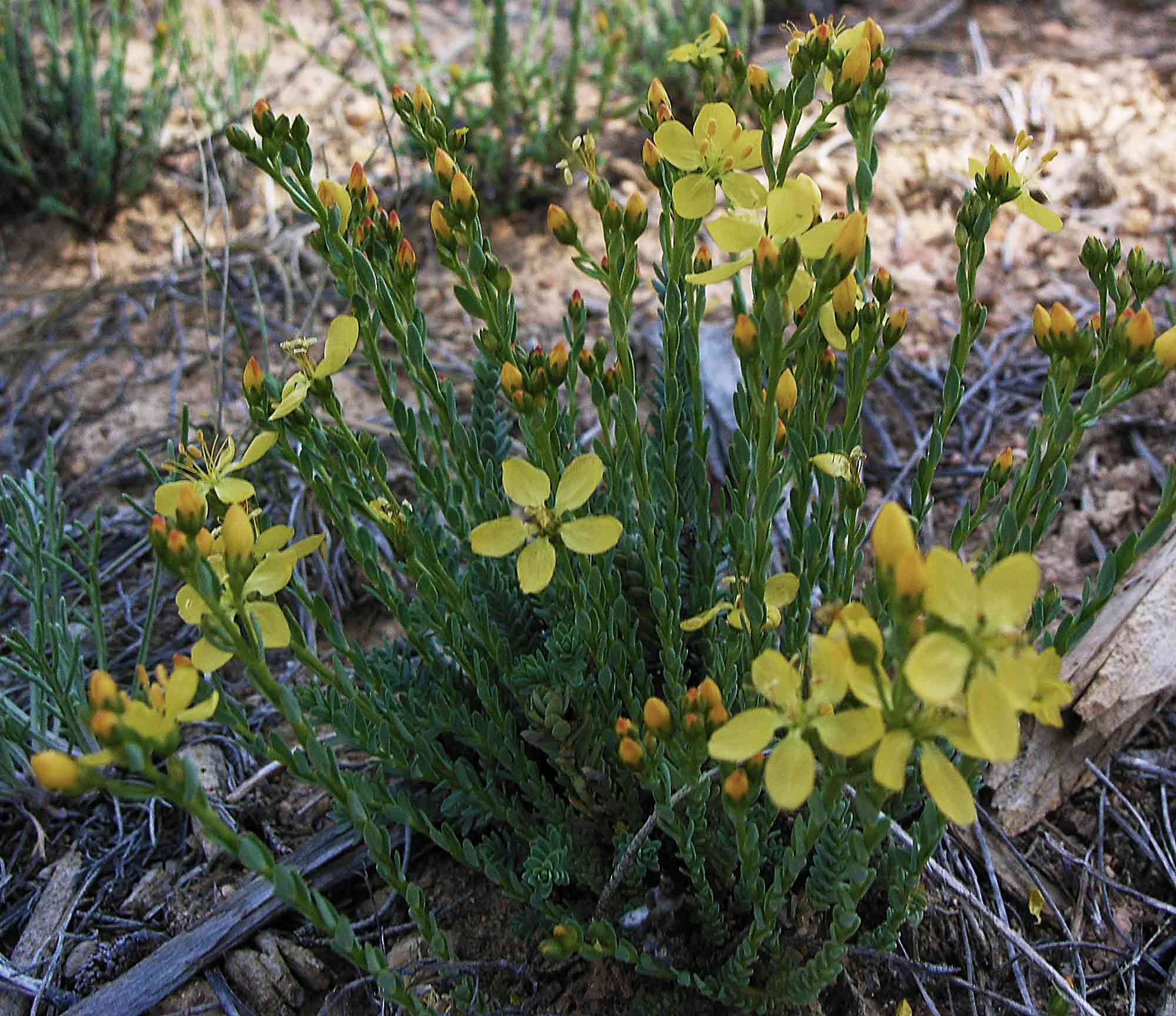
Linum kingii Utah—Great Basin Desert-Utah Plateau. Garfield Co., Dixie NF: Bryce Canyon region, west of the National Park, Red Cap Road; 37º45'02.9", 112º16.11.7", 2151 m. Ponderosa pine oodland. Common. Sample of entire plant (rt-st-lf-fl-fr). Richard Spjut & Paul Burchstead 16377, 25-June-08.
|
|
|
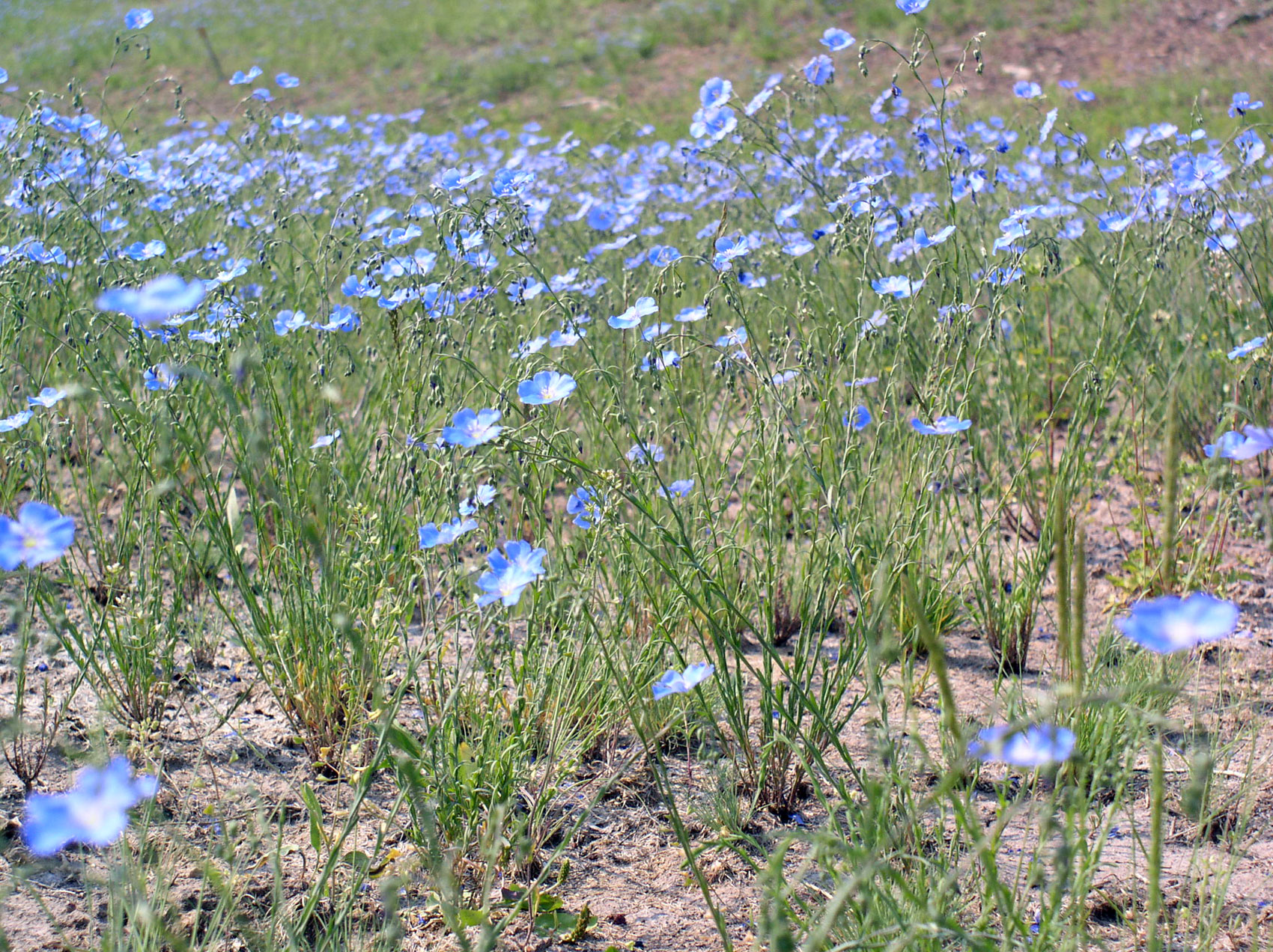
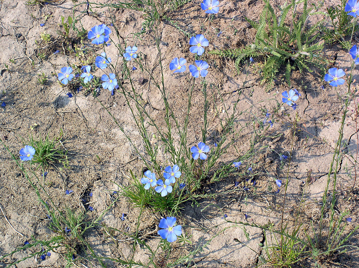
Linum lewisii
|
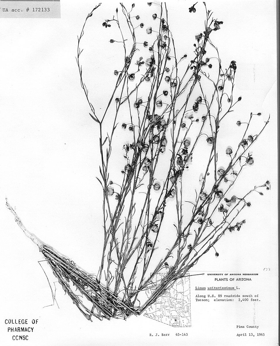
Pima Co., AZ, Barr 65-143 |
|
Beejmohun V., O. Fliniaux, E. Grand, F. Lamblin, L. Bensaddek, P. Christen, J. Kovensky, M. A. Fliniaux and F. Mesnard. 2007. Microwave-assisted extraction of the main phenolic compounds in flaxseed. Phytochem. Anal. 18(4): 275–282. “A microwave-assisted extraction (MAE) method has been applied for the first time to the extraction of the main lignan, secoisolariciresinol diglucoside (SDG), and the two most concentrated hydroxycinnamic acid glucosides in flaxseed. The effects of microwave power, extraction time and alkaline treatment were investigated. It was shown that a 3 min MAE resulted in an SDG content of 16.1+/-0.4 mg/g, a p-coumaric acid glucoside content of 3.7+/-0.2 mg/g and a ferulic acid glucoside content of 4.1+/-0.2 mg/g. These values were compared with those obtained using conventional extraction methods and the results demonstrated that MAE was more effective in terms of both yield and time consumption.” Bergman Jungeström M., L. U. Thompson and C. Dabrosin. 2007. Flaxseed and its lignans inhibit estradiol-induced growth, angiogenesis, and secretion of vascular endothelial growth factor in human breast cancer xenografts in vivo. Clin. Cancer Res. 13(3):1061–1067. “PURPOSE: Vascular endothelial growth factor (VEGF) is a potent stimulator of angiogenesis, which is crucial in cancer progression. We have previously shown that estradiol (E2) increases VEGF in breast cancer. Phytoestrogens are potential compounds in breast cancer prevention and treatment by poorly understood mechanisms. The main phytoestrogens in Western diet are lignans, and flaxseed is a rich source of the mammalian lignans enterodiol and enterolactone. EXPERIMENTAL DESIGN: In the present study, ovariectomized mice were treated with continuous release of E2. MCF-7 tumors were established and mice were fed with basal diet or 10% flaxseed, and two groups that were fed basal diet received daily injections with enterodiol or enterolactone (15 mg/kg body weight). RESULTS: We show that flaxseed, enterodiol, and enterolactone counteracted E2-induced growth and angiogenesis in solid tumors. Extracellular VEGF in vivo, sampled using microdialysis, in all intervention groups was significantly decreased compared with tumors in the basal diet group. Our in vivo findings were confirmed in vitro. By adding enterodiol or enterolactone, E2-induced VEGF secretion in MCF-7 cells decreased significantly without agonistic effects. The increased VEGF secretion by E2 in MCF-7 cells increased the expression of VEGF receptor-2 in umbilical vein endothelial cells, suggesting a proangiogenic effect by E2 by two different mechanisms, both of which were inhibited by the addition of lignans. CONCLUSIONS: Our results suggest that flaxseed and its lignans have potent antiestrogenic effects on estrogen receptor-positive breast cancer and may prove to be beneficial in breast cancer prevention strategies in the future.” Berim A., B. Schneider and M. Petersen M. 2007. Methyl allyl ether formation in plants: novel S-adenosyl L-methionine:coniferyl alcohol 9-O-methyltransferase from suspension cultures of three Linum species. Plant Mol. Biol. 64(3):279-91. “A novel 41 kDa methyltransferase displaying high regiospecificity towards the allylic hydroxyl moiety of coniferyl alcohol was cloned from suspension cultures of Linum nodiflorum L. and expressed in E. coli. The apparent K (m) for coniferyl alcohol is 7.23 microM with a V (max) of 707.5 pkat mg(-1) protein at 30 degrees C, whereas the K (m) for the co-substrate S-adenosyl-L-methionine is 18.5 microM. Structure-function relationship studies revealed stringent structure requirements. Even minor substructure deviations as the side-chain saturation or changes in the phenyl ring substitution result in activities decreased by 75-90%. Crotyl and allyl alcohols are not substrates, confirming that the aromatic ring itself is indispensable, and solely the derivatives with a C(3) side-chain are accepted. The enzyme shares only similarities under 46% on amino acid level with other known methyltransferases. The designated reaction product, coniferyl alcohol 9-methyl ether, could be detected in suspension cells. The highest content of up to 0.02% of the dry mass is concurrent with an increase of the specific enzyme activity that reaches its maximum of 3.94 pkat mg(-1) on day 6 of the culture period. Transcript levels estimated by semi-quantitative RT-PCR remain constant until day 6 and recede thereafter. The corresponding methyltransferase from Linum flavum L. differs mainly by one short variable fragment. Biochemical characterization revealed a higher catalytic efficiency and a slightly broader substrate plasticity together with a lower sensitivity to the presence of Zn(2+), Cu(2+) and Co(2+). This is to our knowledge the first report of a regiospecific allylic O-methylation of phenylpropanoids in plants.” Bommareddy A., B. L. Arasada, D. P. Mathees and C. Dwivedi. 2006. Chemopreventive effects of dietary flaxseed on colon tumor development. Nutr. Cancer 54(2): 216–222. “Fatty acid composition of dietary fat plays a vital role in colon tumor development in animal models. Fats containing omega-6 fatty acids (e.g., corn oil) enhanced and omega-3 fatty acids (e.g., flaxseed oil) reduced chemically induced colon tumor development in rats. Lignans have also been shown to prevent colon tumor development in experimental animals. The objective of this investigation is to study the effects of dietary flaxseed meal, a source of both omega-3 fatty acid and lignans, on colon tumor development and compare them with the effects of dietary corn meal. Male Fischer rats, two groups of 24 each, were assigned to the AIN-93M diet supplemented with either 15% corn meal or 15% flaxseed meal, respectively. Carcinogenesis was initiated with subcutaneous injections of azoxymethane (15 mg/kg) once a week for 3 consecutive wk. After 35 wk of initiation, rats were anesthetized with ether. Blood was collected by cardiac puncture, and rats were sacrificed. The gastrointestinal tract was isolated. The site, size, and number of tumors were recorded. The fatty acid analysis of the collected serum and colon samples was performed. Expression of cyclooxygenase (COX)-1 and COX-2 was performed by Western blot method. Lignan levels in serum and colon samples were assayed. Colon tumor incidence, multiplicity, and size were found to be 82.6% and 29.4%; 1.3 and 0.3; and 44.4 and 5.3 mm(2) in corn and flaxseed meal groups, respectively. Colon and serum samples of the corn meal group showed higher levels of omega-6 fatty acid levels whereas the flaxseed meal group exhibited higher levels of omega-3 fatty acids. COX-1 and COX-2 expression in the flaxseed group was significantly lower (P < 0.05) as compared to the corn group. Dietary flaxseed meal containing high levels of omega-3 fatty acids and lignans is effective in preventing colon tumor development when compared with dietary corn meal possibly by increasing omega-3 fatty acid levels and decreasing COX-1 and COX-2 levels.” Chen J., K. A. Power, J. Mann, A. Cheng and L. U. Thompson. 2007. Dietary flaxseed interaction with tamoxifen induced tumor regression in athymic mice with MCF-7 xenografts by downregulating the expression of estrogen related gene products and signal transduction pathways. Nutr. Cancer. 58(2): 162–170. “Our previous short-term study has shown that 10% flaxseed (FS) inhibits the growth of human estrogen dependent estrogen receptor positive breast tumors (MCF-7) xenografts in ovariectomized (OVX) athymic mice and enhances the tumor inhibitory effect of tamoxifen (TAM). This study determined the long-term effect of 5% and 10% FS, with or without TAM, on the growth of MCF-7 xenografts in athymic mice and the potential mechanisms of actions. OVX mice with established MCF-7 tumors were treated with basal diet (control), 5% FS (5FS), 10% FS (10FS), and TAM (5 mg/pellet, 60-day release), alone or in combination, for 16 wk without estrogen supplementation. Tumor growth was monitored weekly. At sacrifice, the tumors were analyzed by immunohistochemistry for cell proliferation, apoptosis, and expression of estrogen-related genes and signal transduction pathways. Both 5FS and 10FS regressed the pretreatment tumor size by over 90% similar to control. TAM initially regressed the tumors but then induced a regrowth; thus, only a final 6% reduction from pretreatment tumor size was achieved, which was attenuated by combining TAM with 10FS but not with 5FS. TAM combined with 10FS regressed tumors to 55% of pretreatment tumor size due to decreased cell proliferation and increased apoptosis. The expressions of cyclin D1, estrogen receptor alpha, human epidermal growth factor receptor 2, and insulin-like growth factor I receptor in the TAM group were significantly reduced when TAM was combined with 5FS or 10FS. In conclusion, after long-term treatment, FS did not stimulate tumor growth and combined with TAM, regressed tumor size in part due to downregulation of the expression of estrogen-related gene products and signal transduction pathways.” Dupasquier C. M., E. Dibrov, A. L. Kneesh, P. K. Cheung, K. G. Lee, H. K. Alexander, B. K. Yeganeh, M. H. Moghadasian and G. N. Pierce. 2007. Dietary flaxseed inhibits atherosclerosis in the LDL receptor-deficient mouse in part through antiproliferative and anti-inflammatory actions. Am. J. Physiol. Heart Circ. Physiol. 293(4): H2394–402. “Dietary flaxseed has been shown to have potent antiatherogenic effects in rabbits. The purpose of the present study was to investigate the antiatherogenic capacity of flaxseed in an animal model that more closely represents the human atherosclerotic condition, the LDL receptor-deficient mouse (LDLrKO), and to identify the cellular mechanisms for these effects. LDLrKO mice were administered a regular diet (RG), a 10% flaxseed-supplemented diet (FX), or an atherogenic diet containing 2% cholesterol alone (CH) or supplemented with 10% flaxseed (CF), 5% flaxseed (CF5), 1% flaxseed (CF1), or 5% coconut oil (CS) for 24 wk. LDLrKO mice fed a cholesterol-supplemented diet exhibited a rise in plasma cholesterol without a change in triglycerides and an increase in atherosclerotic plaque formation. The CS mice exhibited elevated levels of plasma cholesterol, triglycerides, and saturated fatty acids and an increase in plaque development. Supplementation of the cholesterol-enriched diet with 10% (wt/wt) ground flaxseed lowered plasma cholesterol and saturated fatty acids, increased plasma ALA, and inhibited plaque formation in the aorta and aortic sinus compared with mice fed a diet supplemented with only dietary cholesterol. The expression of proliferating cell nuclear antigen (PCNA) and the inflammatory markers IL-6, mac-3, and VCAM-1 was increased in aortic tissue from CH and CS mice. This expression was significantly reduced or normalized when flaxseed was included in the diet. Our results demonstrate that dietary flaxseed can inhibit atherosclerosis in the LDLrKO mouse through a reduction of circulating cholesterol levels and, at a cellular level, via antiproliferative and anti-inflammatory actions.” Khan G., P. Penttinen, A. Cabanes, A. Foxworth, A. Chezek, K. Mastropole, B. Yu, A. Smeds, T. Halttunen, C. Good, S. Mäkelä and L. Hilakivi-Clarke. 2007. Maternal flaxseed diet during pregnancy or lactation increases female rat offspring's susceptibility to carcinogen-induced mammary tumorigenesis. Reprod. Toxicol. 23(3): 397–406. “Flaxseed contains several dietary components that have been linked to low breast cancer risk; i.e., n-3 polyunsaturated fatty acids (PUFAs), lignans and fiber, but it also contains detectable levels of cadmium, a heavy metal that activates the estrogen receptor (ER). Since estrogenic exposures early in life modify susceptibility to develop breast cancer, we wondered whether maternal dietary intake of 5% or 10% flaxseed during pregnancy or lactation (between postpartum days 5 and 25) might affect 7,12-dimethylbenz[a]anthracene (DMBA)-induced mammary tumorigenesis in the rat offspring. Our data indicated that both in utero and postnatal 5% and 10% flaxseed exposures shortened mammary tumor latency, and 10% flaxseed exposure increased tumor multiplicity, compared to the controls. Further, when assessed in 8-week-old rats, in utero 10% flaxseed exposure increased lobular ER-alpha protein levels, and both in utero and postnatal flaxseed exposures dose-dependently reduced ER-beta protein levels in the terminal end buds (TEBs) lobules and ducts. Exposures to flaxseed did not alter the number of TEBs or affect cell proliferation within the epithelial structures. In a separate group of immature rats that were fed 5% defatted flaxseed diet (flaxseed source different than in the diets fed to pregnant or lactating rats) for 7 days, cadmium exposure through the diet was six-fold higher than allowed for humans by World Health Organization, and cadmium significantly accumulated in the liver and kidneys of the rats. It remains to be determined whether the increased mammary cancer in rats exposed to flaxseed through a maternal diet in utero or lactation was caused by cadmium present in flaxseed, and whether the reduced mammary ER-beta content was causally linked to increased mammary cancer risk among the offspring.” Konuklugil B., I. Ionkova, N. Vasilev, T. J. Schmidt, J. Windhövel, E. Fuss and A. W. Alfermann. 2007. Lignans from Linum species of sections Syllinum and Linum. Nat. Prod. Res. 21(1):1–6. “The aryltetralin lignans 6-methoxypodophyllotoxin, 5'-demethoxy-6-methoxypodophyllotoxin as well as the corresponding 8'-epimers 6-methoxypicropodophyllin, and 5'-demethoxy-6-methoxypicropodophyllin were isolated from suspension cultures of Linum cariense, and 4'-demethyl-6-methoxypodophyllotoxin together with 6-methoxypodophyllotoxin from plants of L. tauricum, which both belong to section Syllinum of the genus Linum. Cell cultures of L. altaicum, L. austriacum ssp. euxinum and L. lewisii belonging to section Linum accumulate the naphthalene lignans justicidin B and isojusticidin B. The different lignans were identified by HPLC and spectroscopic methods.” Mohagheghzadeh A., A. Gholami, S. Hemmati, M. R. Ardakani, T. J. Schmidt and A. W. Alfermann. 2007. Root cultures of Linum species section Syllinum as rich sources of 6-methoxypodophyllotoxin. Z. Naturforsch [C]. 62(1-2): 43–49. “Linum spp. from section Syllinum are promising for the production of aryltetralin lignans like podophyllotoxin (PTOX) and 6-methoxypodophyllotoxin (MPTOX). MPTOX is a PTOX congener that has cytotoxic activity comparable with PTOX. In this study root cultures of Linum bungei from section Dasyllinum, L. strictum from section Linastrum, L. album, L. mucronatum ssp. mucronatum and L. nodiflorum from section Syllinum were established and their MPTOX levels were investigated in 1000 ml flasks. Root cultures of L. mucronatum ssp. mucronatum and L. nodiflorum were used to examine cell growth and production of MPTOX during a culture period of 36 days in 250 ml flasks. Considerable amounts of MPTOX in root cultures (1000 ml flasks) of L. album (6 mg/100 g DW), L. mucronatum ssp. mucronatum (770 mg/100 g DW) and L. nodiflorum (91 mg/100 g DW) were detected while it wasn't detected in root cultures of L. bungei and L. strictum. In time course experiments, the maximum amount of MPTOX in L. nodiflorum root culture was at day 16 with 480 mg/ 100 g DW and the maximum amount of MPTOX in L. mucronatum ssp. mucronatum root culture was at day 12 with 130 mg/100 g DW. The results showed that root cultures of Linum species from section Syllinum are rich sources of MPTOX and since this lignan has remarkable cytotoxic activity, it can be used as a precursor for the production of antitumor agents. Pellizzon M. A., J. T. Billheimer, L. T. Bloedon, P. O. Szapary and D. J. Rader. 2007. Flaxseed reduces plasma cholesterol levels in hypercholesterolemic mouse models. J. Am. Coll. Nutr. 26(1): 66–75. “OBJECTIVE: We examined the effects of whole ground flaxseed added to a Western diet on plasma and hepatic lipids and hepatic gene expression in male and female human apolipoprotein B-100 transgenic (hApoBtg) mice which have a plasma lipid profile more closely resembling man than wild type mice and in mice lacking the low density lipoprotein receptor (LDLr) and apolipoprotein B mRNA editing enzyme complex 1 (LDLr(-/-)/apobec(-/-)). METHODS: The Westernized control diet containing 0.1% cholesterol and 30% kcal as fat was fed for 10 days to hApoBtg mice and for 14 days to LDLr(-/-)/apobec(-/-) mice. Animals from each genetic background were then divided into 2 groups based on gender and mean plasma total cholesterol (TC). The hApoBtg and LDLr(-/-)/apobec(-/-) mice either continued on the control diet for a total of 31 and 35 days, respectively or were fed 20% w/w whole ground flaxseed (flax) with comparable caloric, macronutrient and fiber content for 21 days. Blood was obtained after a 4 hour fast from all mice prior to feeding both control and flax diets, after 10 days on the flax diet, and after 21 days on the flax at which time all mice were exsanguinated. RESULTS: The control diet increased TC by >100 mg/dl in the hApoBtg with a greater increase observed in males and by 800 mg/dl in mice lacking the LDLr. After 3 weeks, the flax diet significantly reduced plasma TC by 19% and 22% in hApoBtg and LDLr(-/-)/apobec(-/-), respectively and non-high density lipoprotein cholesterol (non-HDL-C) by 24% in both models (p for all <0.05). Flax significantly reduced hepatic cholesterol in hApoBtg by 32% and 47% in males and females, respectively and LDLr(-/-)/apobec(-/-) mice by 66%. Flax had no effect on the expression of the following hepatic genes: LDLr, 3-hydroxy-3-methylglutaryl (HMG) CoA reductase, phospholipid transfer protein, cholesterol 7alpha hydroxylase, fatty acid synthase, and acyl CoA oxidase in either mouse model. CONCLUSIONS: Flaxseed reduces plasma and hepatic cholesterol in hApoBtg mice, but had no effect on hepatic lipogenic genes and was equally effective in mice lacking LDLr. The combined data suggest that the lipid lowering effect of flax is not hepatic mediated and may be at the level of cholesterol absorption and/or bile acid reabsorption.” Power K. A. and L. U. Thompson. 2007. Can the combination of flaxseed and its lignans with soy and its isoflavones reduce the growth stimulatory effect of soy and its isoflavones on established breast cancer? Mol. Nutr. Food Res. 51(7): 845–856. “Consumption of phytoestrogen (PE)-rich foods (i. e., soy and flaxseed (FS)) is increasing because of their suggested health benefits. However, recent studies raise concern over the safety of soy and its isoflavones, particularly genistein (GEN), for postmenopausal breast cancer (BC), due to their potential stimulatory effects on human breast tissue and on the growth of existing tumors in rodents. FS, rich in PE lignans, which is metabolized to the mammalian lignans enterolactone (ENL) and enterodiol (END), has consistently been shown to have tumor inhibitory effects in a human clinical trial as well as rodent BC models. Using the preclinical athymic mouse postmenopausal BC model, combining FS with soy protein or GEN with END and ENL, was found to negate the tumor stimulatory effects of soy protein or GEN alone. The mechanism may be related to the modulation of estrogen receptor and MAPK signaling pathways. If these studies can be confirmed in clinical trials, then consumption of combined soy and FS, or their PEs, may reduce the tumor growth stimulatory effect of soy or GEN. This may indicate that if soy is consumed with lignan-rich foods, it may continue to induce its other beneficial health effects, without inducing adverse effect on postmenopausal BC.” Vasilev N., Elfahmi, R. Bos, O. Kayser, G. Momekov, S. Konstantinov and I. Ionkova . 2006. Production of justicidin B, a cytotoxic arylnaphthalene lignan from genetically transformed root cultures of Linum leonii. J. Nat. Prod. 69(7):1014-1017. “Callus and hairy root cultures of Linum leonii were established. The genetic transformation in hairy roots was proven by PCR analysis, which showed integration of rol A and rol C genes into the plant genome. Calli and hairy roots accumulate the arylnaphthalene lignan justicidin B as a major constituent. Hairy roots produce 5-fold higher yields of justicidin B (10.8 mg g(-1) DW) compared to calli. Justicidin B shows strong cytotoxicity on the chronic myeloid leukemia LAMA-8 and K-562 cell lines and on the chronic lymphoid leukemia SKW-3 cell line with IC(50) values of 1.11, 6.08, and 1.62 microM, respectively. Apoptotic properties of justicidin B are reported for the first time.” Vasilev N., G. Momekov, M. Zaharieva, S. Konstantinov, P. Bremner, M. Heinrich and I. Ionkova. 2006. Cytotoxic activity of a podophyllotoxin-like lignan from Linum tauricum Willd. Neoplasma 52(5): 425–429. “The present study describes the preliminary evaluation of the cytotoxic activity of a podophyllotoxin-like compound 4'-demethyl-6-methoxypodophyllotoxin (4'-DM-6-Mptox), isolated as one of the main lignans of Linum tauricum Willd. ssp. tauricum. The cytotoxic effects 4'-DM-6-Mptox were assessed by the MTT-dye reduction assay against the human leukemic cell lines HL-60, BV-173 and LAMA-84. DNA-fragmentation analysis and NF-kB inhibition assay were performed in order to elucidate some of the mechanistic aspects of the cytotoxic action of the investigated compound. 4'-DM-6-Mptox was found to exert prominent cytotoxicity, with IC50 values being several-fold lower than those of the referent antineoplastic agent etoposide. The DNA-fragmentation analysis revealed that 4'-DM-6-Mptox treatment triggered apoptosis in BV-173 and HL-60 cells. In our hands 4'-DM-6-Mptox was found to induce concentration-dependent NF-kB inhibition in HeLa cells as assessed by the IL-6 luciferase gene reporter assay, which though not quite prominent, at least partly contributes to the cytotoxic potential of the tested lignan. On the basis of the results obtained it could be concluded that 4'-DM-6-Mptox necessitates further pharmacological and toxicological evaluation as a possible chemotherapeutic agent. Furthermore due to its relatively high concentrations in the described plant source the possibility for its use as a precursor for the semisynthetic production of lignan-based drugs, could be considered.”
|
|